Showing 18 items matching x-ray tube
-
 Federation University Historical Collection
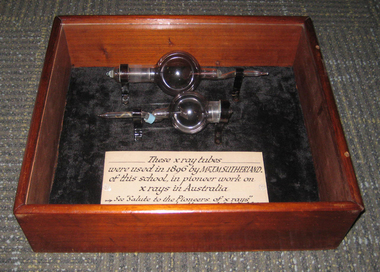
Equipment - Object, Early X-Ray Tube, 1896, c1896
... Early X-Ray Tube, 1896...Two X-ray tubes used in pioneering x-rays at the Ballarat... Equipment Early X-Ray Tube, 1896 Two X-ray tubes used in pioneering ...Pioneer xrays were conducted at the Ballarat School of Mines in 1896.Two X-ray tubes used in pioneering x-rays at the Ballarat School of Mines in 1896. They have been boxed for safety. x-ray, xray, xray tube, john m. sutherland, ballarat school of mines, xray demonstration, pioneer xrays -
 The Ed Muirhead Physics Museum
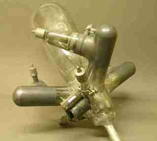
Gas X-ray Tube
... Gas X-ray Tube...gas x-ray tube.... For this particular experiment Lyle actually made his own x-ray tube. His... Tube The investigation of the x-ray appears early on to have ...The investigation of the x-ray appears early on to have been a priority research topic at the University of Melbourne’s School of Physics. This interest was sparked by the appointment in 1889 of Professor T.R. Lyle. Lyle, who was head of the school until 1915, is thought to have been the first person in Australia to have taken an x-ray photograph. A copy of this photograph can be found in the School of Physics Archive. For this particular experiment Lyle actually made his own x-ray tube. His successor, Professor Laby, continued to work with x-rays. During the 1920s Laby worked on the x-ray spectra of atoms and in 1930 he co-published with Dr. C.E. Eddy, Quantitative Analysis by X-Ray Spectroscopy. Also with Eddy, Laby produced the landmark paper Sensitivity of Atomic Analysis by X-rays. Laby went on to have an x-ray spectrograph of his own design manufactured by Adam Hilger Ltd. (see cat. No. 38). School of Physics, the University of Melbourne Cat. No. 22. Jacqueline Eager Student Projects Placement, Cultural Collections 2005 The original X-ray tubes relied on low pressure operation. The electrons and positive ions are produced in the residual gas. Positive ions are accelerated towards the cathode and release electrons which on hitting the anode produce X-rays. These early gas X-ray tubes operated satisfactory only over a narrow pressure range. Stamped Label: “NATURAL PHILOSOPHY LABORATORY/ No/ UNIVERSITY OF MELBOURNE” Stamped: “90268 M. No. 5171[??]/No. 2156[??]/ M. No. 346585.” x-ray tubes, gas x-ray tube, laby, spectroscopy -
 Federation University Historical Collection
Scientific Instument, Hewittic Rectifiers, X-Ray Tube, 1956
... X-Ray Tube...x-ray tube... Scientific Instument X-Ray Tube Mercury arc rectifier, 3-phase input ...This item was acquired by the Ballarat School of Mines Electrical Engineering department for use in electric power laboratory as a source of D.C., and also for instructional purposes. This central mercury arc element was located in a cabinet with transparent side panels, and equipped with the required electric accessories, to be a self-contained stand-alone unit. Head of the Electrical Engineering Department at the time was John M. Sutherland.Mercury arc rectifier, 3-phase input. Constructed of blown glass, and complicated configuration: the central inverted truncated cone is provided with 3 large diameter "horns' and four smaller ones. Each horn has electrical connection to outside, some have side horns. Approximately half a cup of free mercury inside the glass complex. No. 33369scientific instrument, x-ray, x-ray tube, xray, john m. sutherland, electrical engineering, ballarat school of mines -
 Federation University Historical Collection
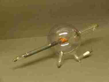
Scientific Instument, X-Ray Tube
... X-Ray Tube...x-ray tube... Scientific Instument X-Ray Tube A spherical glass vessel with two ...A spherical glass vessel with two principles electrodes mounted inside opposing cylindrical extensions. A third mounted in an offset extension. A fourth electrical connection in a branched extension, perpendicular to main axis. scientific instrument, x-ray tube, xray, physics -
The Ed Muirhead Physics Museum
X-ray Tube, Machlett
... X-ray Tube, Machlett ...This X-ray tube was designed to provide electrostatic...Glass bulbous X-ray tube attached at either end to metal... Streets The University of Melbourne Parkville melbourne X-ray Tube ...This X-ray tube was designed to provide electrostatic protection for the filament (cathode) so as to permit long life to be achieved at operating voltages in the range 100-300kV. It is not certain whether the tube was in use within the School by Professor Laby’s X-ray group or whether it was presented to the School by a medical user. It would be somewhat surprising if it fitted into this School of Physics Research Program at a date as late as 1933 when tubes with demountable anodes were in use.Glass bulbous X-ray tube attached at either end to metal electrodes. Mounted for demonstration on a wooden base. Dated1937On glass bulb: “Machlett Patent 1954016” -
The Ed Muirhead Physics Museum
Gas X-ray Tube, Victor
... Gas X-ray Tube, Victor.... For this particular experiment Lyle actually made his own x-ray tube. His... Tube, Victor The investigation of the x-ray appears early ...The investigation of the x-ray appears early on to have been a priority research topic at the University of Melbourne’s School of Physics. This interest was sparked by the appointment in 1889 of Professor T.R. Lyle. Lyle, who was head of the school until 1915, is thought to have been the first person in Australia to have taken an x-ray photograph. A copy of this photograph can be found in the School of Physics Archive. For this particular experiment Lyle actually made his own x-ray tube. His successor, Professor Laby, continued to work with x-rays. During the 1920s Laby worked on the x-ray spectra of atoms and in 1930 he co-published with Dr. C.E. Eddy, Quantitative Analysis by X-Ray Spectroscopy. Also with Eddy, Laby produced the landmark paper Sensitivity of Atomic Analysis by X-rays. Laby went on to have an x-ray spectrograph of his own design manufactured by Adam Hilger Ltd. (see cat. No. 38). School of Physics, the University of Melbourne Cat. No. 22. Jacqueline Eager Student Projects Placement, Cultural Collections 2005 The original X-ray tubes relied on low pressure operation. The electrons and positive ions are produced in the residual gas. Positive ions are accelerated towards the cathode and release electrons which on hitting the anode produce X-rays. These early gas X-ray tubes operated satisfactory only over a narrow pressure range. Manufacturer’s mark stamped: “PATENTED/ VICTOR/ TRADEMARK/ MADE IN BOSTON U.S.A./ TUNGSTEN” A white circular stamp, stamped near the manufacturer’s mark: “[illegible]TER WIGGH[illegible]” Stamped label: “NAT. PHIL. LAB./ No./ UNIV. OF MELB.” Inscription on the end face of the copper piece: “PAT. SEP 5’ 11 DEC. 30’13/ JUNE 23, 14 NOV. 30.15/ 43835” -
The Ed Muirhead Physics Museum
Coolidge X-ray Tube
... Coolidge X-ray Tube.... For this particular experiment Lyle actually made his own x-ray tube. His...-ray Tube The investigation of the x-ray appears early ...The investigation of the x-ray appears early on to have been a priority research topic at the University of Melbourne’s School of Physics. This interest was sparked by the appointment in 1889 of Professor T.R. Lyle. Lyle, who was head of the school until 1915, is thought to have been the first person in Australia to have taken an x-ray photograph. A copy of this photograph can be found in the School of Physics Archive. For this particular experiment Lyle actually made his own x-ray tube. His successor, Professor Laby, continued to work with x-rays. During the 1920s Laby worked on the x-ray spectra of atoms and in 1930 he co-published with Dr. C.E. Eddy, Quantitative Analysis by X-Ray Spectroscopy. Also with Eddy, Laby produced the landmark paper Sensitivity of Atomic Analysis by X-rays. Laby went on to have an x-ray spectrograph of his own design manufactured by Adam Hilger Ltd. (see cat. No. 38). School of Physics, the University of Melbourne Cat. No. 22. Jacqueline Eager Student Projects Placement, Cultural Collections 2005 In 1913 Coolidge overcame the limitation of the narrow operating range of the gas X-ray tubes with the invention of the vacuum X-ray tube. A filament heated by an electric current directly releases electrons by thermionic emission. In thermionic emission, electrons are emitted from a metal surface directly by the application of an electric current to heat a wire filament. The electrons accelerate to the anode and produce X-rays. The anode has associated cooling fins due to the high temperatures attained by the release of kinetic energy by the electrons on colliding with the anode. Internal Glass sleeve: “A941/L2593/2821” -
The Ed Muirhead Physics Museum
Rotating Anode X-ray Tube
... Rotating Anode X-ray Tube.... For this particular experiment Lyle actually made his own x-ray tube. His... Anode X-ray Tube The investigation of the x-ray appears early ...The investigation of the x-ray appears early on to have been a priority research topic at the University of Melbourne’s School of Physics. This interest was sparked by the appointment in 1889 of Professor T.R. Lyle. Lyle, who was head of the school until 1915, is thought to have been the first person in Australia to have taken an x-ray photograph. A copy of this photograph can be found in the School of Physics Archive. For this particular experiment Lyle actually made his own x-ray tube. His successor, Professor Laby, continued to work with x-rays. During the 1920s Laby worked on the x-ray spectra of atoms and in 1930 he co-published with Dr. C.E. Eddy, Quantitative Analysis by X-Ray Spectroscopy. Also with Eddy, Laby produced the landmark paper Sensitivity of Atomic Analysis by X-rays. Laby went on to have an x-ray spectrograph of his own design manufactured by Adam Hilger Ltd. (see cat. No. 38). School of Physics, the University of Melbourne Cat. No. 22. Jacqueline Eager Student Projects Placement, Cultural Collections 2005 A modern X-ray tube differs little from the original Coolidge tube. A minor modification is the rotating anode type that extends the life and increases the available power of the tube by presenting a new portion of the anode when required. “P125/20/40/NrF038803 (?) SIEMENS-REINIGER-WERRE AG ERLANGEN Eigen filleung (?) mind. 0,7 mm AL” On rotating shaft: “FO/33803” On cathode: “23C” -
 University of Melbourne, School of Chemistry
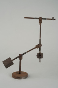
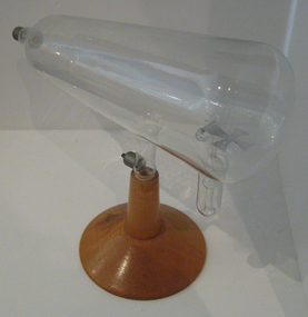
Vacuum Tube Stand
... Stand for Vacuum or X-Ray Tubes.... for Vacuum or X-Ray Tubes. ...Stand for Vacuum or X-Ray Tubes. -
 Federation University Historical Collection
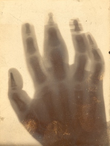
Photograph - X-ray, X-Ray of a boys hand, c1896
... xray x-ray roentgen tube ...X-ray experiments were conducted at the Ballarat School of Mines in July 1896, with a public demonstration on 22 July 1896. J.M. Sutherland pioneered works on X-rays in Australia by conducting displays in Ballarat in 1896.Photograph of an X-ray of a four year old boy's diseased second finger, mounted on card."Boy 4 yrs old diseased 2nd finger"ballarat school of mines, j.m.sutherland, pioneer x-ray, xray, x-ray, roentgen tube -
 Federation University Historical Collection
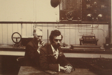
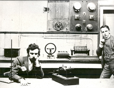
Photograph - Black and White, X-Ray Demonstration at the Ballarat School of Mines, 1896
Four months after the discovery of the Roentgen tube the Ballarat School of Mines were conducting pioneer X-ray demonstrations in Ballarat. This photograph is thought to be taken at that time, and is framed along with some of the x-rays taken at that time. Digital copy of two men sit in front of x-ray equipment at the Ballarat School of Mines. The man in the foreground is John McKenzie Sutherland. The copy is from the original framed 1896 X-Rays (Cat. No. 1243)ballarat school of mines, x-ray, xray, scientific instruments, john sutherland, sutherland, photography, foto, roentgen tube, john m. sutherland, john sutherland -
 Federation University Historical Collection
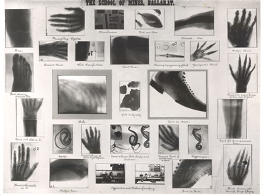
Photograph - Original x-rays, X-rays from pioneering Ballarat Demonstrations, 1896
X-Rays were first discovered on 08 November 1895. By 18 July 1896 staff members of the Ballarat School of Mines (SMB), were experimenting with the exciting new discovery. The history of x-rays began on 08 November 1895 at the University of Wurzburg in Bavaria. The discovery was officially announced on 25 December 1895. The first radiographs in Ballarat were taken at the School of Mines in July 1896 according to the Ballarat School of Mines (SMB) Annual Report. Frederick J. Martell, the Registrar of SMB arranged for the importation of tubes, while John M. Sutherland, an electrician, conducted most of the experiments giving 6 inch, 12 inch and 16 inch sparks respectively. In a short time brilliantly successful results were obtained, with some SMB Roentgen negatives taken at this time still in existence today. Samuel Ernest Figgis, H. R. W. Murphy, D. McDougall, and Frederick J. Martell carried out experiments at the SMB on Saturday evening 18 July 1896, producing 'perfect' negatives of a hand and wrist. A Roentgen Tube and an induction coil giving a two inch spark, the coil being sparked by the SMB's dynamo, were used to obtain these results. The Courier reported that 'the exposure of five minutes was ample' but concluded that 'the length of the exposure will be shortened as experiments proceed.' The Ballarat Courier reported on 20 July 1896 that: "Thanks to the energy of the staff of The School of Mines, Ballarat, and particularly to Messers F.J. Martell and D. McDougall, the assistance of Rontgen X-rays will soon be available, for the relief of suffering humanity, at this institution." Martell was an ardent amateur photographer, and Duncan McDougall's experience as an electrician has enabled the two gentlemen to carry their experiments on to a perfectly successful issue. At first these gentlemen, together with Professor Purdie and Mr W. Huey Steele, conducted a series of experiments by the aid of a Bonetti glass-plate induction machine which had been constructed by Mr McDougall. The results were very good, the various bones of the hand being distinctly visible. The following people were among those who witnessed the first X-ray experiments to be carried out in Ballarat. Andrew Anderson, President of the School of Mines, a large number of ladies and gentlemen, the medical profession Dr Edward Champion (1867-1929) Dr Gerald Eugene Cussen (1888-1943) Dr William Edward Davies (1868-1928) Dr Charles William Henry hardy (1861-1941) Dr Edward Kenneth Herring(1864-1922) Dr Joseph Lalor (1859-1907) Dr James Thomas Mitchell (1856-1945) Dr Edward Graham Ochiltree (1857-1896) Dr Robert Denham Pinnock (1849-1902) Dr Joseph Francis Usher (c1832-1909) Dr Grace Vale (nk-1933). The staff of SMB Professor Alfred Mica Smith Professor D. J. Dawbarn Mr. F. J. Martell. (http://guerin.ballarat.edu.au/aasp/is/library/collections/art_history/honour-roll/honourroll_X-Ray_pioneers.shtml) "Seven patients of the doctors who were present were treated. In each case the patient was suffering from the effects of an old wound or some other injury to other a hand or foot. One patient, a boy had a finger broken some years earlier and when xray plate of the injury was developed it indicated plainly the fracture on one of the joints of the third finger. In another case a woman had a foot had a foot X-rayed and the photograph revealed the seat of an earlier imjury to it. Likewise an injury to the bones of a hand of an elderly man was revealed by an x-ray photograph. Every X-ray photograph that evening showed the importance of this new development in electrical science and the doctors present agreed the "the results of the experiments on their patients, by showing the exact seat of the injury and its present condittion, would materially assist them iin supporting cures." (Warren Perry, The Ballarat School of Mines and Industries Ballarat, p 146.)A number of photographic xrays as taken by the Ballarat School of Mines in 1896. The xrays include bones, hands, feet, shoes and more. ballarat school of mines, scientific equipment, x-ray, xray, photography, foto, roentgen tube -
 Federation University Historical Collection
Equipment - Scientific Instrument, Crookes Tube
... discovered. Wilhelm Röntgen discovered X-rays using the Crookes tube ...A Crookes tube is an early experimental electrical discharge tube, invented by English physicist William Crookes and others around 1869-1875, in which cathode rays, streams of electrons, were discovered. Wilhelm Röntgen discovered X-rays using the Crookes tube in 1895. The term is also used for the first generation, cold cathode X-ray tubes, which evolved from the experimental Crookes tubes and were used until about 1920.(http://en.wikipedia.org/wiki/Crookes_tube)Glass vacuum tube with a metal maltese cross inside. The tube is on a timber stand.scientific instruments, crookes tube, science, maltese cross, scientific objects -
 Federation University Historical Collection
Photograph - Photograph - Black and White, X-Ray demonstration at the Ballarat School of Mines, 1896
X-Rays were first discovered on 8 November 1895. By 18 July 1896 staff members of the Ballarat School of Mines (SMB), were experimenting with the exciting new discovery. The history of x-rays began on 8 November 1895 at the University of Wurzburg in Bavaria. The discovery was officially announced on 25 December 1895. The first radiographs in Ballarat were taken at the School of Mines in July 1896 according to the Ballarat School of Mines (SMB) Annual Report. Frederick J. Martell, the Registrar of SMB arranged for the importation of tubes, while John M. Sutherland, an electrician, conducted most of the experiments giving 6 inch, 12 inch and 16 inch sparks respectively. In a short time brilliantly successful results were obtained, with some SMB Roentgen negatives taken at this time still in existence today. Samuel Ernest Figgis, H. R. W. Murphy, D. McDougall, and Frederick J. Martell carried out experiments at the SMB on Saturday evening 18 July 1896, producing 'perfect' negatives of a hand and wrist. A Roentgen Tube and an induction coil giving a two inch spark, the coil being sparked by the SMB's dynamo, were used to obtain these results. The Courier reported that 'the exposure of five minutes was ample' but concluded that 'the length of the exposure will be shortened as experiments proceed.' The Ballarat Courier reported on 20 July 1896 that: "Thanks to the energy of the staff of The School of Mines, Ballarat, and particularly to Messers F.J. Martell and D. McDougall, the assistance of Rontgen X-rays will soon be available, for the relief of suffering humanity, at this institution." Martell was an ardent amateur photographer, and Duncan McDougall's experience as an electrician has enabled the two gentlemen to carry their experiments on to a perfectly successful issue. At first these gentlemen, together with Professor Purdie and Mr W. Huey Steele, conducted a series of experiments by the aid of a Bonetti glass-plate induction machine which had been constructed by Mr McDougall. The results were very good, the various bones of the hand being distinctly visible. The following people were among those who witnessed the first X-ray experiments to be carried out in Ballarat. Andrew Anderson, President of the School of Mines, a large number of ladies and gentlemen, the medical profession Dr Edward Champion (1867-1929) Dr Gerald Eugene Cussen (1888-1943) Dr William Edward Davies (1868-1928) Dr Charles William Henry hardy (1861-1941) Dr Edward Kenneth Herring(1864-1922) Dr Joseph Lalor (1859-1907) Dr James Thomas Mitchell (1856-1945) Dr Edward Graham Ochiltree (1857-1896) Dr Robert Denham Pinnock (1849-1902) Dr Joseph Francis Usher (c1832-1909) Dr Grace Vale (nk-1933). The staff of SMB Professor Alfred Mica Smith Professor D. J. Dawbarn Mr. F. J. Martell. (http://guerin.ballarat.edu.au/aasp/is/library/collections/art_history/honour-roll/honourroll_X-Ray_pioneers.shtml)Two men, one seated, one standing standing in a room with early electrical equipment. They are conducting pioneer X-rays at the Ballarat School of Mines in 1896. Seated on the left is John Waters Sutherland.A bromide of this images states "Experimenting with X-rays, 1896"ballarat school of mines, x-ray, xray, john sutherland, sutherland, electricity, photography, foto, roentgen tube, medical -
Federation University Historical Collection
Correspondence, Letters from A. Gallenkamp & Co to the Ballarat School of Mines, 1898
... -ray xray rontgen tubes crooks tubes uranium tubes ...Two hand written letters relating to orders from the Ballarat School of Mines .1) 31 March 1898 Dear Sir, In reply to your favor of 23 February we have nearly all the apparatus now ready you were good enough to order. I've [illegible] here 12 cases [illegible] fomr Robinson's goods we likely had to wait about 5 months for supply from works, the demand having been quite extraordinary this year. Ou people [illegible] are so busy with Rontgen ray tubes that we cannot get crokes ad Uranium tubes as quickly as we should like. Yours are not yet to hand, have however a promise of delivery very shortly when all goods will be ready for dispatch. Now we have [illegible] in our very large new warehouses in Sun Street, we are importing [illegible] and other glass goods in four or five times the quantity, we could store in our old place, so that we shall hold presently the August stock of glass apparatus in London, enabling us to fill orders of every size in much shorter issue. We shall not loose a day or a book so soon as we are ready with the order early in April. We beg to call your attention to fene glass listed by the [illegible] book post. Your esteemed further orders will oblige. Yours truly [Illegible] .2) 09 September 1898 Dear Sir We thank you for the favor of your order of August just received and give [illegible] attention. arriving to our ... For Reagent bottle 1757 you state no size, we will give you 250cm as the size usually order here. ... We beg to confirm our letter of 2nd inst and enclose two circulars of interest. Yours Faithfully [illegible]a. gallenkamp and co., letterhead, ballarat school of mines, frederick martell, x-ray, xray, rontgen tubes, crooks tubes, uranium tubes -
The Ed Muirhead Physics Museum
Lyle Radiograph, 3/3/1896
"Salute to the X Ray Pioneers of Australia" by W Watson &Sons Ltd [1946] [Medical Library 610.9 WAT page 24-27 radiograph reproduction p26]Copies of the Lyle radiograph (see below) are on file with the letter (9Sept 1982) from J F Richardson (Australian Radiation Laboratory, as it was then called) detailing the description of the reproduction as follows: RADIOGRAPH OF PROFESSOR ORME MASSON'S FOOT! MOST PROBABLY THE FIRST RADIOGRAPH TAKEN IN AUSTRALIA. TAKEN BY PROFESSOR LYLE ON MARCH 3RD, 1896 USING A CROOKES DISCHARGE TUBE OF HIS OWN CONSTRUCTION -
Federation University Historical Collection
Book, Alfred P. Morgan (revised by J.W. Sim), The Boy Electrician, 1941 (exact); George G. Harrap (publisher)
Graham Beanland, and his father, C.H. Beanland, both attended the Ballarat School of Mines.Red hard covered book of 328 pages. Contents include: Magnets and magnetism, static electricity, cells and batteries, cables, measuring, telegraphs, telephones, microphones, coils, transmission, transformers, generators, electric motors, radio, electric railways, lighting, circuits, gas-discharge tubes, Tesla coil, copper wire.Book Plate: Yallourn Technical School. Presented to G. Beanland. Second Prize Form I. Dec 1942 C.H. Beanland, pricipallighting, telephones, ammeter, magnets, magnetisn, static electricity, cells, batteries, cables, measuring, telegraphs, microphones, coils, transmission, transformers, generators, electric motors, radio, electric railways, circuits, gas dischatge tubes, tesla coil, copperiwire, morse code, galvanometer, television, ohm, x ray, xray, graham beanland, c h beanland, charles beanland, yallourn -
 Kiewa Valley Historical Society
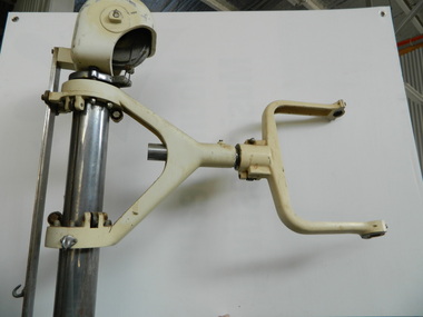
Medical Equipment - X-ray Machine
This tall piece of equipment was used in the theatre room of the Tawonga District Hospital to move (by rolling it) to the bedside of the patient and then to adjust the large light over the area of operation as required by the surgeon.The Tawonga District Hospital's theatre room was well equipped with up to date technical equipment.Stainless steel metal tube attached to 4 legs of steel that spray out into a 'star' and have a roller coaster attached at their end. On the 5th end of the star a steel arm comes up and has a tray attached to its end. At the top of the cylinder is attached a cream metal opened ended cap with a wheel which has thick wire around it. Below this top and on the cylinder a cream metal arm comes out like a spanner, the end of which a large light could be fitted. This arm can be moved up and down the tube. This stand also has a control 'radiation control unit' that can be attached to it and to the power.medical equipment. operation. surgeon. tawonga district hospital. theatre room., x-ray, falls creek medical centre